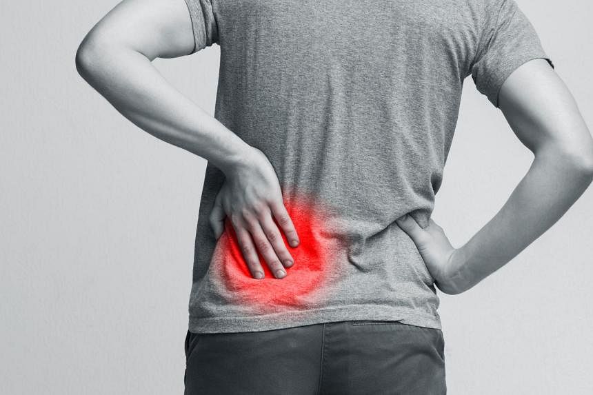
Experiencing lower back pain or a burning sensation when urinating? It may be a sign of kidney stones.
This common urological condition happens when there is a high concentration of certain minerals and chemicals in the patient’s urine, leading to the formation of crystals.
“The stones are tiny when they first form and gradually increase in size as more minerals are deposited,” says Dr Koh Li-Tsa, a senior urologist from Urohealth Medical Clinic.
It affects approximately 10 per cent of Singaporeans, and while kidney stones can occur at any age, they tend to form in adults over the age of 30. Men are slightly at higher risk than women for developing the condition because they tend to drink less water and consume more protein and salt in their diet, notes Dr Koh.
Other risk factors include a diet high in protein, salt and sugar, excessive sweating, insufficient water intake, obesity, family or personal history of kidney stones, certain digestive or metabolic disorders, and consumption of certain medications, adds Dr Koh.
Diagnosing kidney stones and choosing a suitable procedure
In some cases, a tiny stone can be passed out from your urine without any issue.
“However, kidney stones can get stuck in the ureter and obstruct the flow of urine, causing severe pain. They can even cause the kidney to swell up, get infected and this may lead to kidney failure,” says Dr Koh.
“So, it’s important to have the stones promptly removed to preserve kidney function.”
Patients with lower abdomen and back pain, blood in the urine, and other urinary symptoms such as an urge to urinate or cloudy and foul-smelling urine, are encouraged to undergo tests to determine if the cause is a kidney stone, says Dr Koh. Nausea and vomiting, fever and/or chills are other symptoms to look out for.

To diagnose a kidney stone, you will undergo an X-ray called a "KUB X-ray'' (kidney-ureter-bladder X-ray) which will indicate the size of the stone and its position. This is usually done with a kidney and bladder ultrasound to rule out other possibilities such as obstruction of the urinary tract and kidney tumours.
“Treatment of the stone depends on size, location, and whether it is causing obstruction or pain. The first step of drinking large amounts of water generally works best if the stone is small and located in the lower ureter,” says Dr Koh.
“Should any medication be given, it is usually alpha-blockers, which is an off-label use, but commonly given to relax the ureteric muscles as a form of medical expulsion therapy.”
If the stone is too large, or blocks the flow of urine, or if there is a sign of infection, surgery may be your only solution.
The good news is that you no longer have to endure two to three hours of open surgery with recovery and downtime. Today, there are three minimally invasive treatments that offer effective relief with fewer complications.
1. A 20-minute shock wave treatment that breaks down small stones
Extracorporeal shock wave lithotripsy (ESWL) uses shock waves to break a large stone into smaller pieces so that they can travel more easily out of the body. Patients have to ensure they do not move so that the shockwaves will land on the targeted stone.
Dr Koh says, “It may sound intense, but the entire procedure takes about only 20 minutes and has been proven to be effective on stones smaller than 2cm and those formed in the kidney or ureter.
“Patients may be sedated during ESWL as the shockwaves can be painful, especially at higher levels of intensity. If you are awake, you may be given medication to reduce the pain.”

Another added benefit: You can return to your normal activities on the same day. However, Dr Koh does advise against exercise and strenuous activities for at least a week following the procedure.
“ESWL is not suitable for pregnant women, patients with bleeding disorders and those with large aortic aneurysms (dilatation of a major vessel in the abdomen). It should also not be performed without first treating any urinary tract infection or obstruction of the urinary tract below the level of the stone,” adds Dr Koh.
2. Inserting a tiny scope to directly extract the stone
Ureteroscopy involves using a tiny scope to enter the ureter and kidney. Urohealth senior urologist Dr Tricia Kuo explains: “The scope contains a small device or laser that breaks down the stones into smaller pieces. The fragments are then removed using basket extraction.”
Ureteroscopy allows the doctor to visualise and have direct contact with the stone, so the procedure has a slightly higher success rate compared with ESWL, says Dr Kuo.
“We are also able to identify and treat other possible problems, such as narrowing of the ureter,” she adds.
Since doctors can use scopes of various length, diameter and flexibility, this procedure can be done to treat stones in any part of the urinary tract and for stones of most sizes.
“If the stones are particularly large, they may be challenging to remove using this technique alone. In such cases, alternative treatment options may be explored,” says Dr Kuo.
Ureteroscopy can be done under general or regional anaesthesia, and depending on where the stone is located, the surgery can take anywhere between 30 minutes and two hours.
While you can return to your normal activities on the same day, Dr Kuo advises patients to take it easy with exercise and strenuous activities for at least a week.
Ureteroscopy is not suitable for patients with severe structural abnormalities or deformities in the urinary tract, such as extensive scarring or a previous surgery, as it may be difficult to navigate the scope through the ureter or reach the target area. Pregnant women are also discouraged from opting for this procedure.
3. Accessing the stone through a small incision
If you have bigger stones – larger than 2cm in diameter – Percutaneous nephrolithotomy (PCNL) may be your best option.
It involves making a minor 1cm to 2 cm incision on your back and inserting small telescopes and instruments to access the kidney.
“Pneumatic devices and/or lasers are then used to break up or remove the kidney stone. Although there is an incision, it is still smaller than traditional open kidney surgery and considered minimally invasive,” notes Dr Kuo.
The procedure is done under general anaesthesia and can last for up to three hours. You will also be required to stay in the hospital for one to three days post-surgery.
“During this time, the medical team monitors the patient's condition, manages pain, and allows the initial healing to take place,” says Dr Kuo.
PCNL is unsuitable for pregnant women, patients with severe bleeding disorders or if they have risks associated with anaesthesia.
Ultimately, early detection and response is key to maintaining your kidney health, say both Dr Kuo and Dr Koh, who advise patients to speak to their doctors for the best course of treatment.


